02 Scientific software · 3D morphology
Organoid Explorer
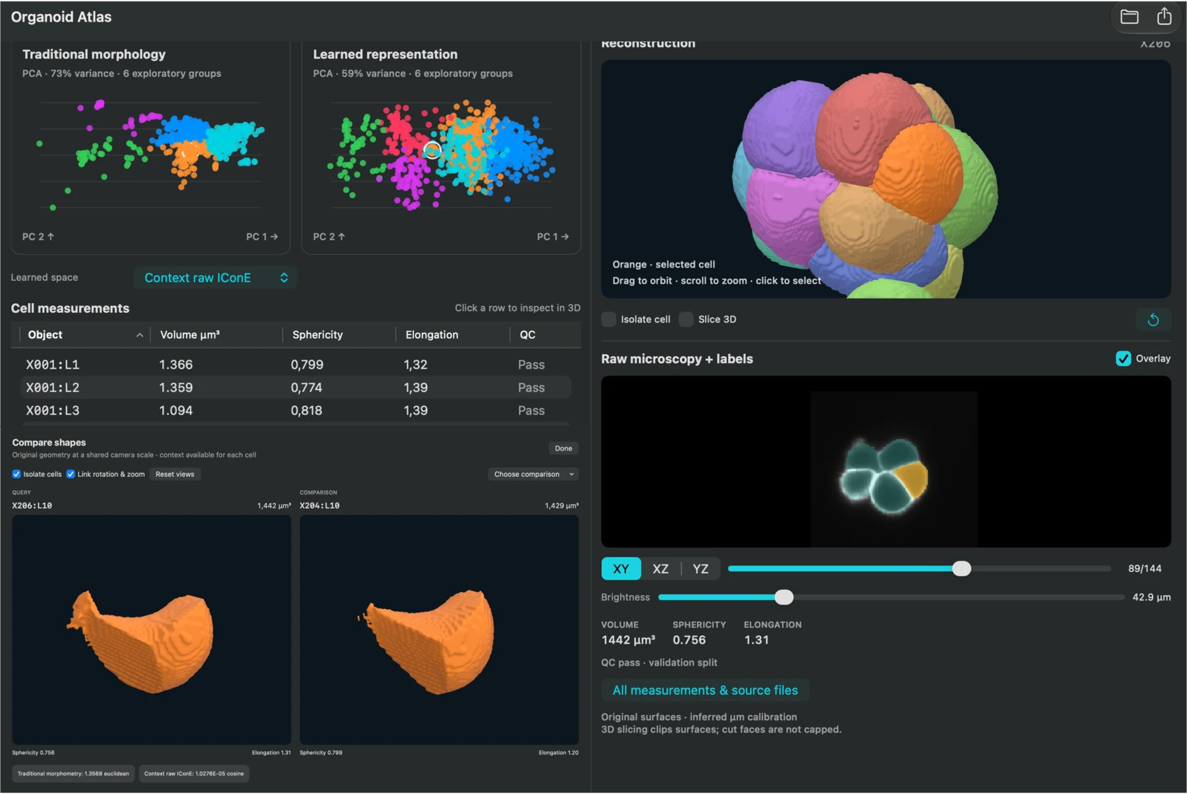
Which cells have similar 3D shapes? This macOS app, published as Organoid Atlas, links quantitative shape comparisons to reconstructed surfaces and the original microscopy.
Motivation
What makes two cells similar?
Volume and sphericity are useful summaries, but cells with similar measurements can have very different boundaries. I wanted a way to compare surfaces directly and trace each one back to the microscopy it came from.
Implementation
Link shape back to the image
The project reconstructed 1,088 cells across 108 microscopy stacks. The viewer connects each cell’s source slices and 3D surface with measurements, embeddings, and nearest-neighbor comparisons.
Project animation
From organoid to shape atlas
Watch the reconstructed cells separate, move into a map of shapes, and reveal a query cell’s nearest matches. The motion is illustrative and the cell surfaces are reconstructed from the project’s microscopy data.
Check a comparison against the source
Linked views make it possible to select a cell in a plot and inspect its reconstructed surface and raw slices. That connection helps distinguish a genuine boundary feature from a segmentation artifact.